Mouse brain region thought to lack a body map has one, found in its resting activity
Columbia researchers traced a hidden layout in the secondary motor cortex that responds most when several body parts move together.
 By Hannah Bell
· 5 min read
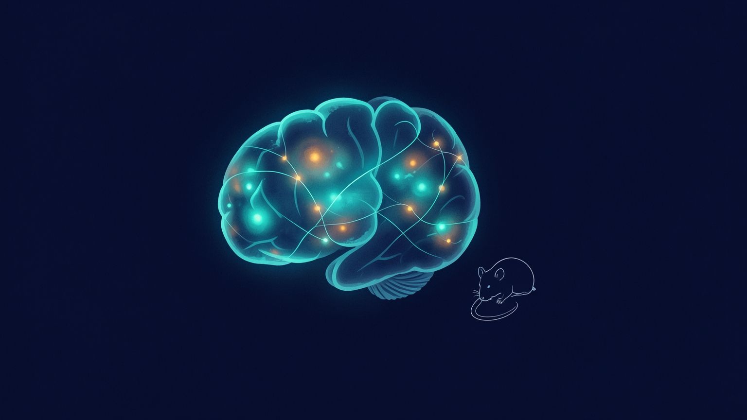
A part of the mouse brain that neuroscientists long believed had no orderly map of the body turns out to have one. Columbia University researchers found it by studying what the brain does when the animal is sitting still.
The study, published in Nature Communications, describes a body-part-specific layout in the secondary motor cortex, or M2, of awake mice. Separate patches of M2 correspond to separate body parts, such as the forepaws and the mouth. Those same patches fired hardest when a mouse used several body parts at once, for instance raising both forepaws to its mouth and licking them.
That second result may matter more than the map itself. Picking up a cup and bringing it to your lips takes coordinated work from the shoulder, hand, head and mouth. Scientists have struggled to explain how the brain links those motions into one fluid act. A region that is organized by body part and becomes most active during multi-part movements is a strong candidate for part of the answer. The work was led by Weihao Xu, who did the research as a doctoral student and later a postdoctoral scientist in the lab of Elizabeth Hillman at Columbia's Zuckerman Mind Brain Behavior Institute.
How resting activity gave the map away
The brain's main movement area, the primary motor cortex or M1, has been known for decades to contain a body map. Stimulate one spot and a specific finger or limb twitches. That stimulation method is the source of the familiar "homunculus" pictures of the human brain, with outsized hands and lips draped across the cortex. Applied to M2, the same approach produced murky results. Many researchers concluded that M2 had no such layout and handled movement in more abstract terms. Earlier work had tied M2 to complex combinations of motions, but its activity had been hard to read in terms of particular body parts.
Mice make that problem harder. Even a well-trained mouse rarely moves one body part on its own, so it is difficult to link a patch of cortex to a single limb by watching behavior. The Columbia team used high-speed wide-field optical imaging, developed in Hillman's lab, to record activity across the surface of the cortex as mice moved. The more revealing signal, the researchers said, came from the apparently random activity of the brain at rest.
In that resting activity, small spots in M2 rose and fell in step with matching regions of the sensory cortex. The sensory cortex has a well-established body map, so each synchronized M2 spot could be labeled with a body part. Assembled together, Xu said, the spots formed "a body map hidden in the intrinsic chatter of M2." He called M2 "a part of the brain that wasn't supposed to have a body map."
Xu spent three years checking the result. The obvious worry was that the signal was spilling over from M1 next door. To rule that out, the team used two-color wide-field optical mapping to separate corticospinal neurons from other cells. Corticospinal neurons send commands down the spinal cord and cluster most heavily in M1. The synchronized fluctuations were not prominent in those neurons, which the authors read as evidence that the patterns are not motor output and that the map sits in M2. Anatomical tracing then showed that each sensory region sends connections to its matching M2 subregion. The paper also reports that the resting fluctuations were strongest at rest and died down once the animals moved.
Finally, the researchers watched M2 during spontaneous movement. The patches lit up for the body parts the map predicted, and the response was stronger when several parts worked together.
"Confusingly, M2 chatters when the body is still and cares more about complicated movements than simple ones." said Hillman, the study's corresponding author.
Hillman, now chair of the Department of Imaging Sciences at St. Jude Children's Research Hospital, said those traits explain why M2's activity had been so hard to interpret. That is a fair diagnosis of the earlier confusion. Electrical stimulation asks a region to produce a simple movement. A region that cares more about combinations than single twitches would naturally give unclear answers to that test.
What the mouse result can and cannot tell us
This is a study of mice, and the findings describe mouse anatomy and activity. Rodent M2 is often compared with premotor and supplementary motor areas in primates. Still, the paper does not show that the human brain holds the same hidden map or that it works the same way.
The evidence linking M2 to coordination is also correlational. The researchers recorded which patches became active during multi-part movements and found stronger engagement for coordinated actions. The reports do not describe experiments that silenced or disrupted those patches to test whether the movements depend on them. Until someone runs that kind of test, "M2 coordinates complex movement" remains a well-supported hypothesis rather than a demonstrated mechanism. The movements studied were also spontaneous ones, not trained tasks designed to isolate coordination.
The medical and engineering payoffs are the researchers' hopes, not results. Xu said the map could help scientists understand the dexterous movements people can lose after strokes and other brain injuries. Hillman expects the work to help explain how the brain plans and adjusts movements from moment to moment, with possible relevance to movement disorders and to building better robots. Xu also put the engineering challenge bluntly, describing coordinated movement as "so complex that robots still struggle with it." These are reasonable directions, but this mouse imaging study offers no stroke treatment and no robot design.
The method may prove as useful as the map. Neuroscientists studying people already rely heavily on resting-state brain scans, which look at which regions fluctuate together when a person is doing nothing in particular. The Columbia study adds a concrete case in which resting synchrony pointed to a structure that stimulation had missed. The team then confirmed that structure with wiring and behavior. Xu said he hoped other researchers would build on the idea of using "how the brain's resting activity can reveal its hidden wiring."
Funding came from National Institute of Neurological Disorders and Stroke and National Institute of Mental Health grants under the BRAIN Initiative. Other support came from the Simons Collaboration on the Global Brain, Foundation Leducq and ALSAC, the fundraising and awareness organization of St. Jude. Xu has since left academia and now works as a data scientist in industry. He credited his computer science and machine learning training with making the analysis possible.
The next test is the one that would move this from map to mechanism. If silencing a patch of M2 disrupts a coordinated movement but leaves its single-part pieces intact, the case for M2 as a coordination hub would be hard to argue with.
Sources
Hannah Bell
Hannah Bell writes about physics, experimental discoveries, and the ideas used to describe the physical world. Her coverage explores how researchers test theories, interpret measurements, and investigate unresolved questions. She makes unfamiliar concepts approachable while distinguishing what an experiment demonstrates from the possibilities it may open for future research.
View all stories