Two brain regions shrink before the hippocampus in early Alzheimer's, study finds
Johns Hopkins researchers say the entorhinal cortex and amygdala lose tissue first, possibly a decade before symptoms. A screening test is still years away.
 By Amara Okafor
· 5 min read
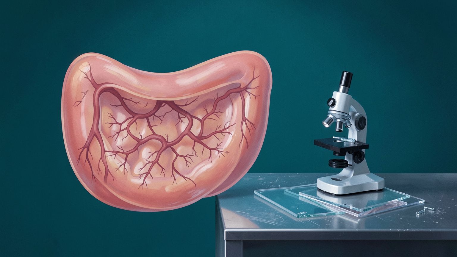
Two small brain structures next to the hippocampus appear to lose tissue earlier than the hippocampus itself as Alzheimer's disease takes hold. Johns Hopkins University researchers reached that conclusion after analyzing hundreds of brain scans from people followed over time.
The regions are the entorhinal cortex and the amygdala. Both showed greater relative shrinkage in the disease's early stages than the hippocampus, the memory center doctors have traditionally examined for signs of Alzheimer's damage. The team says this kind of atrophy can show up as much as 10 years before memory problems do. The work was published in the journal NeuroImage Reports and is being presented at Neuroscience 2026, the Society for Neuroscience's annual meeting.
"It's at least a 5-to-10-year play where your brain is changing," said Michael Miller, a biomedical engineer and the study's senior author. He argued that treating the disease a few years into that window, instead of after memory symptoms appear, is the real prize. "That's the great promise here for your loved ones who are at risk."
That promise is worth taking seriously, and it is also worth keeping in proportion. The study points to candidate early markers. It does not deliver a test that tells a healthy 45-year-old whether Alzheimer's is coming. The researchers make that distinction themselves.
The team drew on MRI and PET scans from two independent research efforts, the Alzheimer's Disease Neuroimaging Initiative and the Biomarkers of Cognitive Decline Among Normal Individuals study. Participants spanned the course of the disease. Some had no symptoms, some had cognitive impairment, and some eventually died of Alzheimer's.
The same pattern turned up in both groups: early on, the entorhinal cortex and amygdala lost proportionally more tissue than the hippocampus. Seeing the result hold across two separate cohorts is the most convincing part of the work. A finding drawn from a single dataset can reflect quirks in how that group was recruited or scanned. A finding that repeats in a second one is harder to dismiss.
The two regions do different jobs. The entorhinal cortex helps the brain retrieve memories it has already stored. The amygdala shapes emotional memory, meaning how emotionally charged experiences are processed and recalled. The researchers' argument is that tracking both structures together could catch early changes that are easy to miss when attention stays fixed on the hippocampus.
The researchers also tied the shrinkage to tau, one of the two proteins most closely linked to Alzheimer's. In healthy nerve cells, tau helps hold the cell's internal scaffolding together. In Alzheimer's it builds up abnormally and forms tangles that disrupt normal function. Using PET imaging and digital pathology, the team found that areas where gray matter had thinned overlapped with areas showing tau-related changes. Gray matter is the brain tissue packed with nerve cell bodies.
That overlap fits a long-standing picture from pathology. Researchers have described tau tangles appearing early in and around the entorhinal region for decades, so finding measurable tissue loss there first makes biological sense. Still, overlap is an association. It strengthens the case that shrinkage in these regions reflects the disease process, but it does not by itself show how much of the tissue loss tau causes.
Co-author Meaghan Morris, a neuropathologist who studies Alzheimer's and other dementias, framed the payoff as better sorting of patients by risk. As both biomarkers and treatments become more widely available, she said, "knowing who is going to have cognitive decline is going to be really important." The goal, she added, is to treat people earlier and slow the disease's course.
Marilyn Albert, a co-author who directs the Johns Hopkins Alzheimer's Disease Research Center, described the larger aim in similar terms: treating people before symptoms emerge so the disease can be delayed or even stopped.
The researchers believe measuring tissue loss in both regions, rather than leaning mainly on the hippocampus, could eventually improve predictions of who will decline. They envision a future in which people with a family history of Alzheimer's or other risk factors might have these regions examined as early as their 30s or 40s, especially as imaging becomes cheaper and easier to get.
That vision is a long way from routine care, and several gaps stand between here and there. The study materials released so far give no participant counts and no measures of predictive accuracy, such as how often the pattern correctly flags someone who later declines. Those numbers will matter far more to doctors than the overall pattern.
Measurement is another hurdle. The entorhinal cortex and amygdala are small, and detecting a modest relative loss of tissue usually requires comparing scans over time. A single snapshot may not be enough. Screening healthy middle-aged adults would also raise practical questions about cost, access and what to tell people whose scans look worrisome before any symptom exists. The researchers acknowledge that more work is needed before these changes can serve as a reliable diagnostic tool.
Earlier warning signs are only as useful as what doctors can do with them. That is why the team's next step points toward biology rather than screening. The researchers plan to study what happens inside these regions at a much finer scale, mapping the molecular changes that surround abnormal tau buildup and drive tissue loss. Miller calls these areas molecular neighborhoods, and he sees them as the route to new treatments.
"Once we know more about the molecular neighborhoods, we can develop therapies," he said.
That ordering is the right one. A scan that reveals Alzheimer's a decade early would matter most to patients if it arrived alongside a therapy built to act during that decade.
Sources
Amara Okafor
Amara Okafor covers community health, access to services, and developments affecting everyday wellbeing. Her work follows research and public information about the conditions that influence health across different communities. She focuses on explaining how services are organized, whom new initiatives may reach, and what the available evidence says about their effects.
View all stories